


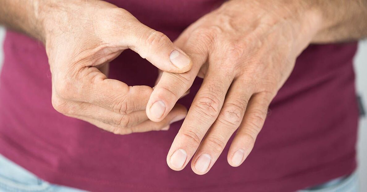
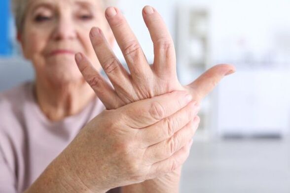
Pain in the fingersoccurs when bones, joints, soft tissues, blood vessels, nerves are affected. It can be dull, acute, weak, intense, permanent, intermittent, short-term. It is often associated with motor activity, weather conditions, and other factors. Simultaneous external disturbances are possible: deformities, color and temperature changes, edema. The results of a survey, external examination, x-ray and other methods are used to determine the cause of finger pain. Until the diagnosis is made, rest is recommended, sometimes taking painkillers.
Why do my fingers hurt
Traumatic injuries
Finger injury is characterized by moderate pain. Then the intensity of the pain gradually decreases. Edema, hyperaemia, cyanosis, bleeding are possible. The function of the finger is slightly impaired. Hematomas on the palm surface of the fingers are manifested by moderate pain, skin peeling, and the formation of a cavity filled with dark blood. In subungual hematomas, the pain is intense, twitching, pulsating, aggravated by lowering the brush. Partial or complete detachment of the nail plate is possible.
Finger fracture is accompanied by severe, explosive pain at the time of injury. After that, the pain decreases somewhat but remains intense. The finger turns blue, swells, and its functions are severely impaired. Demonstrable deformity, crepitus, abnormal mobility. When a finger moves, sharp pain is observed. The finger is deformed, swelling when it tries to move in the affected joint, spring resistance is determined.
In the first hours of freezing, the pain is mild, tingling. Then the pain syndrome intensifies and gets a burning character. The finger swells and becomes cyanotic. In case of deep freezing, there is no sensitivity in the distal parts, the fingers are cold, pale, there is pain at the border of healthy and affected tissues.
Infectious lesions
Panaritium is characterized by rapidly increasing pain, swelling, hyperemia, cyanosis, and abscess formation. Pain twitching, throbbing, depriving sleep at night. Subungual panaritium and deep forms of the disease (bone, joint, tendon) are particularly painful. In the superficial forms of panaritium (skin, periungual, subcutaneous, subungual) the general condition is mild, with deep symptoms of poisoning and fever.
Chinga is formed in people who cut and process the carcasses of marine wildlife, with minor injuries: abrasions, wounds, cracks. It occurs as a dull, mild pain in the wound area, which is replaced 1-2 days later by pain in the joint of the finger (usually the proximal). The pain grows, it becomes aching, throbbing, supplemented by swelling, pallor and cyanosis of the finger.
Arthritis
Finger joint pain in rheumatoid arthritis is symmetrical. Grade 1 activity is indicated by less joint pain and transient stiffness. In grade 2, the pain is disturbed at rest and during movement, combined with prolonged stiffness, limited mobility, redness. Grade 3 is characterized by intense constant pain, persistent stiffness, swelling, hyperemia. Movement is severely restricted.
Gout of the fingers is more common in women. One or more joints may be affected. Pain is usually acute, acute, combined with edema, hyperemia, impaired function, and general fever. Canceled symptoms are less common - mild pain and mild redness with a satisfactory general condition.
Psoriatic arthritis occurs suddenly or gradually. In the first case the pain is moderate, increasing, in the second - sharp, intense. At the peak of the disease, the characteristic picture is: pain, worsens at night and at rest, weakens during the day, with movements, swelling of the fingers, purple-blue discoloration of the skin. The distal interphalangeal joints are most commonly affected. Over time, multiple deformations appear.
In post-traumatic arthritis, one joint is involved. Infectious-allergic forms of the disease underlying bacterial and viral infections are characterized by multiple lesions. In the case of professional peripheral arthritis, the most stressed joints of the fingers are involved in the process. Pain in each of the listed forms of pathology intensifies at night, weakens during the day, and is accompanied by morning stiffness, local swelling, and difficulty moving. Deformations are observed during long runs.
Degenerative pathologies
With arthrosis of the hand, the pain is initially indeterminate, intermittent, short-lived. There is morning stiffness. After that, the painful feelings intensify, prolong, sometimes burning, can be perceived with any movement, restrict daily activities, and perform delicate operations. Heberden and Bouchard junctions are created. Lateral deformations are formed.
Diseases of the ligaments and tendons
Patients with narrowing ligamentitis are concerned about the pain along the surface of the palm of the affected finger. At first, the pain syndrome occurs only with pressure and small movements, and then remains at rest. Movements are restricted, accompanied by a click. Over time, flexion contracture develops, and after clicking, there is pain in the arm.
In the initial stage, de Quervain’s disease manifests itself in pain during abduction, protrusion of the first finger. This is followed by aching, depressing pain during any physical activity, with some patients disturbed even at rest. Typical irradiation of the distal phalanx or forearm from the side of the first finger.
Angiotroponeurosis
Raynaud's syndrome is caused by vasospasm accompanied by paroxysmal numbness in the cold fingers. The pain occurs in the second phase of the attack, combined with a breaking, burning sensation and a feeling of fullness. Pain syndrome is short-lived, replaced by a feeling of heat, redness in the distant parts of the hands. The pathology occurs in a number of diseases of various origins, including:
- rheumatoid arthritis;
- systemic lupus erythematosus;
- scleroderma;
- Sharp syndrome;
- anti-synthetase syndrome;
- upper extremity thromboangiitis obliterans;
- endocrine, metabolic, occupational pathologies.
In the absence of other diseases that provoke this condition, Raynaud’s disease is described as having similar pain syndrome. This form is more common in women.
Erythromelalgia occurs on its own or in patients with endocrine, neurological, haematological diseases. This is manifested in paroxysmal attacks of frying, burning pain, edema, hyperemia of the fingers. It is possible for pain to spread from one limb to another, or to occur in a region of both limbs at the same time. The attacks of pain are so strong that they interfere with movement. The pain decreases as the hand cools and raises, and increases as the hand warms up and lowers.
Neurological pathologies
Finger pain occurs when the nerves are damaged, spread in the innervation zone, are of a shooting or burning nature, and are accompanied by sensory disturbances and autonomic-trophic abnormalities. Possible neurological causes:
- Neuropathy of the central nerve.The pain in the I-III. it is localized on the palm side of the fingers and is coupled with the inability to bend the fingers, pinch the hand into the fist, and contrast the finger I.
- carpal tunnel syndrome.Median is a type of neural neuropathy caused by compression of nerve fibers at the wrist level. Localization of pain - as in the previous case. Typical nocturnal seizures, reduction of pain when lowering arms, shaking brushes.
- Radial nerve neuropathy.In the lesion at the level of the forearm and wrist, pain is observed on the back surface of the first finger and the hand, sometimes spreading to the second and third fingers. Irradiation of the forearm and numbness of the hand are typical.
- Neuropathy of the ulnar nerve.The pain is mainly localized in the area of the elbow joint, but can radiate to the hands, IV-V fingers. Pain syndrome often worsens in the morning.
Tumors
Benign tumors affecting the bones of the fingers include chondromas and osteoid osteomas. Chondromas manifest as non-intense pain sensations, the localization of which is unclear, osteoid osteomas - sharp pain in the affected area. Malignant neoplasm of the fingers is rare.
Other reasons
Finger and hand pain is seen in patients with writing cramps, which develops along with occupational neurosis, some other mental and neurological disorders. Pain occurs while writing, working on a computer or typewriter. They break, pull, with tremors, sudden hand weakness, accompanied by local cramps. In addition, finger pain can be seen in the following pathologies:
- Leukemia: Waldenström macroglobulinemia.
- Tumors of the adrenal glands: aldosteroma.
- Complications of diabetes: diabetic neuropathy.
- Vascular diseases: distal digital embolism due to occlusion of the subclavian artery.
- hereditary diseases: Fabry disease.
- Childhood diseases: neuro-arthritis diathesis.
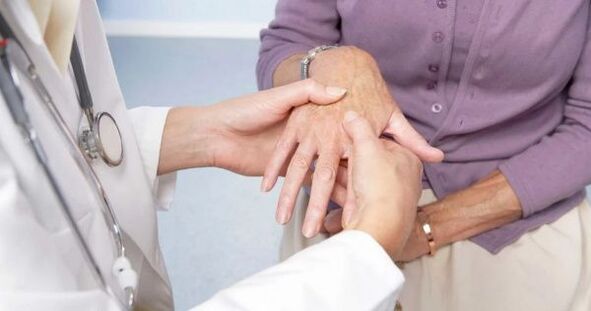
Diagnostics
Traumatologists-orthopedists work to determine the causes of finger pain. The diagnosis is made on the basis of the conversation with the patient, external examination data and further examinations. The diagnostic program includes:
- Survey. The doctor finds out when and under what circumstances the pain syndrome and other symptoms first appeared, determines the characteristics of the dynamics of the development of the disease, the factors that cause the patient to improve or worsen. Study of life history, family history.
- Physical analysis. The specialist will assess the appearance of the fingers, revealing deformities, inflammation, cracks, dry skin, temperature and discoloration, swelling, and other manifestations of the pathology. Investigate sensitivity, range of motion, pulsation in peripheral arteries.
- Radiography.It is performed in two projections, gripping the affected fingers or the whole hand. It reinforces areas of fractures, dislocations, tumors, inflammatory and degenerative processes, destruction of solid structures in deep panaritium forms.
- Electrophysiological examinations.In the case of pain of neurological origin, it is performed to clarify the extent of nerve damage, to assess the condition of the muscles, and to guide the nerves.
- Laboratory tests. It is used to determine inflammation, to assess the general condition of the body, and to detect specific markers of collagenoses.
Patients are referred to an endocrinologist, neurologist, vascular surgeon, and other specialist for consultation. Assignment of CT, MRI, other instrumental techniques. Perform a biopsy of the hard and soft structures for cytological or histological examination.
Treatment
First aid
In case of traumatic injuries, a cold, elevated position of the limb is recommended. The hands are secured with rails or impromptu materials (such as planks). The brush is lifted or a scarf is used. With intense pain syndrome, painkillers are given, and chloroethyl is used in the absence of external damage.
Help for illness is determined by the nature of the pathology - changes in the position of the limb, warming or, conversely, cooling can help. The most common measure is rest, however, in some diseases (carpal tunnel syndrome, arthritis) the pain syndrome is reduced while motor activity is maintained. Acute twitching pain, pronounced signs of inflammation, general hyperthermia warrant urgent consultation with a specialist.
Conservative therapy
In case of displacements and fractures, local anesthesia, reduction and plaster casting are performed. Conservative treatment of traumatic and non-traumatic pathologies of the fingers involves the following activities:
- Protective mode. They are selected according to the nature and severity of the disease. Possible suggestions for limiting the load, using orthopedic devices, applying a plaster bandage.
- Medical therapy. Non-steroidal anti-inflammatory drugs, antibiotics, drugs that improve blood circulation and neurotropic drugs are used. Blockages are indicated with corticosteroids.
- Non-pharmacological methods. Practice therapy, massage, physiotherapy, manual therapy, kinesiotaping are prescribed.
Surgical procedures
Surgeries are performed when conservative methods are ineffective to reduce the duration of treatment and improve long-term outcomes. Taking into account the characteristics of the lesion, the following should be done:
- Injuries: fractures, fixation of dislocations with a knitting needle, necrectomy, amputation of fingers in case of freezing.
- Infectious diseases: opening of panaritium, drainage, occasionally amputation or disarticulation in case of severe lesions.
- Diseases of tendons and ligaments: dissection of the dorsal ligament and excision of adhesions in de Quervain’s disease, dissection of the annular ligaments in constricted ligamentitis.
- Neoplasms: removal of neoplasia, bone resection.
- Neurological diseases: nerve decompression.
Antibiotic therapy is prescribed after surgery. Patients undergo comprehensive rehabilitation aimed at maximum restoration of hand function.